What Is Pectus Carinatum Brace Treatment?
19/10/2026
What Is the Nuss Procedure? How Is It Performed?
19/10/2026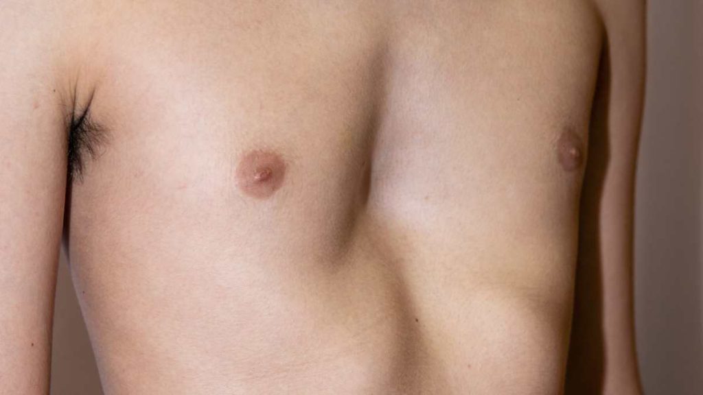
Pectus excavatum is a chest wall deformity in which the breastbone and the front of the chest appear sunken inward. It is sometimes referred to as “funnel chest.”
In some people, the deformity is mild and mainly affects appearance. In more pronounced cases, the shape of the chest may influence the position of the heart, cardiac filling, and the space available for the lungs to expand.
However, the extent to which pectus excavatum affects the heart or lungs cannot be determined from appearance alone.
Evaluation may include the depth and shape of the deformity, the patient’s age, symptoms, exercise capacity, and, when necessary, imaging and functional tests.
What Is Pectus Excavatum?
Pectus excavatum is a chest wall deformity in which the sternum and adjacent costal cartilages are displaced inward.
The depression may be:
- central,
- more pronounced in the lower sternum,
- more prominent on one side,
- symmetrical,
- asymmetrical.
In some children, the deformity is visible from an early age. In others, it becomes more noticeable during periods of rapid growth, particularly during adolescence.
Is Pectus Excavatum Only a Cosmetic Problem?
Not in every patient.
Mild pectus excavatum may cause no measurable functional impairment and may mainly represent a difference in chest appearance.
In some moderate or severe cases, however, changes in chest geometry may influence the mechanical environment of the heart and lungs.
It is therefore not accurate to consider every case purely cosmetic.
At the same time, it is also incorrect to assume that every person with pectus excavatum has impaired heart or lung function.
How Can Pectus Excavatum Affect the Heart?
The heart lies behind the sternum between the two lungs.
When the sternum is displaced backward in pectus excavatum, the amount of space within the chest can be reduced.
In more pronounced deformities, the heart may:
- shift toward the left side,
- rotate within the chest,
- experience compression, particularly on the right-sided chambers.
The degree of these anatomical changes varies considerably between patients.
Can Pectus Excavatum Compress the Heart?
In some patients with a deep deformity, the reduced distance between the sternum and spine can create external pressure on the heart.
The right side of the heart lies relatively close to the sternum and may therefore be more affected in certain cases.
However, visible contact or compression on imaging does not automatically mean that the heart is functioning abnormally.
The clinical significance depends on symptoms and, when needed, additional cardiac evaluation.
Can Pectus Excavatum Affect Heart Function?
In selected patients with more severe deformity, mechanical restriction may affect the way the heart fills with blood.
During physical activity, the heart needs to increase the amount of blood it pumps to meet the body’s increased demands.
If the available space restricts cardiac filling, the ability to increase cardiac output during exercise may be reduced in some patients.
Possible symptoms may include:
- reduced exercise tolerance,
- early fatigue,
- shortness of breath,
- palpitations.
These symptoms are not specific to pectus excavatum, so other possible causes should also be considered.
Can Pectus Excavatum Cause Palpitations?
Some patients with pectus excavatum report palpitations.
Changes in heart position or mechanical compression may contribute to an increased awareness of heartbeat in certain individuals.
However, palpitations may also be caused by:
- heart rhythm disorders,
- thyroid disease,
- anemia,
- stress,
- caffeine,
- other cardiac conditions.
For this reason, palpitations should not automatically be attributed to pectus excavatum.
Electrocardiography or rhythm monitoring may be considered when clinically appropriate.
How Can Pectus Excavatum Affect the Lungs?
The lungs expand as the chest cavity enlarges during breathing.
In pectus excavatum, the reduced front-to-back diameter of the chest may limit available intrathoracic space in more pronounced cases.
This effect is not the same in every patient.
Some people may have normal or near-normal lung function at rest, while limitations become more noticeable during physical activity.
Can Pectus Excavatum Cause Shortness of Breath?
Yes, some patients report shortness of breath, especially during exertion.
It may become more noticeable during:
- running,
- climbing stairs,
- intense exercise,
- prolonged physical activity.
However, shortness of breath can also result from asthma, low physical conditioning, heart disease, lung disease, anemia, and many other conditions.
The symptom should therefore be evaluated in context.
Can Pectus Excavatum Reduce Lung Capacity?
Some patients with more severe pectus excavatum may show reduced lung volumes on pulmonary function testing.
This may be related to mechanical restriction of chest wall expansion.
However, the severity of the external deformity does not always correlate directly with pulmonary function.
Some patients with a visually pronounced deformity may have normal respiratory tests, while others with a less dramatic appearance may experience limitations during exercise.
Why Can Symptoms Be More Noticeable During Exercise?
At rest, the cardiovascular and respiratory systems operate at relatively low demand.
During exercise:
- the heart must pump more blood,
- breathing rate increases,
- lung ventilation increases,
- oxygen delivery to the muscles rises.
If chest wall anatomy creates a mechanical limitation, the effect may become more evident during exercise than at rest.
This is why some patients feel comfortable during ordinary daily activities but notice reduced endurance during sports.
Why Do Some Patients Tire More Easily?
Reduced exercise tolerance may have several contributing factors.
In a patient with pectus excavatum, these may include:
- limitation of cardiac filling during exercise,
- reduced chest wall expansion,
- lower lung volumes,
- poor physical conditioning,
- posture-related mechanics.
In other patients, no significant physiological impairment may be found.
Does Pectus Excavatum Prevent Exercise or Sports?
Pectus excavatum does not automatically prevent a person from exercising.
Many patients with mild and asymptomatic deformity can participate in normal physical activity.
However, further evaluation may be appropriate if exercise causes:
- significant shortness of breath,
- chest pain,
- dizziness,
- fainting,
- unusual palpitations,
- marked reduction in performance compared with peers.
Any exercise restrictions should be based on the individual patient rather than the appearance of the chest alone.
How Is the Effect on the Heart and Lungs Evaluated?
Assessment is not based solely on visual examination.
Depending on the patient’s symptoms and clinical findings, evaluation may include imaging and functional tests.
Why Is CT Used in Pectus Excavatum?
Computed tomography can provide detailed information about chest wall anatomy.
It may help assess:
- depth of the deformity,
- internal chest dimensions,
- position of the heart,
- possible cardiac compression,
- symmetry or asymmetry of the chest.
However, CT is not necessary for every patient.
In children and adolescents, the need for imaging is considered carefully because of radiation exposure.
What Is the Haller Index?
The Haller Index is a radiological measurement commonly used to describe the anatomical severity of pectus excavatum.
It is calculated by dividing the widest internal transverse diameter of the chest by the shortest distance between the sternum and the spine.
As the chest becomes more compressed from front to back, the Haller Index generally increases.
However, the Haller Index should not be used alone to decide whether surgery is necessary.
Other factors include:
- symptoms,
- cardiac compression,
- respiratory findings,
- exercise tolerance,
- chest wall anatomy,
- impact on quality of life.
What Is the Correction Index?
The Correction Index is another radiological measurement used to assess the depth of pectus excavatum.
It estimates the percentage of chest depression compared with the expected anterior-posterior chest dimension.
In some patients, it can provide additional anatomical information alongside the Haller Index.
Like the Haller Index, it does not determine treatment by itself.
Why Is Echocardiography Used?
Echocardiography is an ultrasound examination of the heart.
When clinically indicated, it can help assess:
- cardiac chamber anatomy,
- heart valves,
- pumping function,
- right-sided heart structures,
- possible effects of external compression.
It may be particularly useful in patients with significant deformity or cardiac symptoms.
Is an ECG Needed?
An electrocardiogram records the electrical activity of the heart.
It may be used in patients with palpitations, suspected rhythm disturbances, or certain cardiac symptoms.
However, there is no rule that every patient with pectus excavatum requires an ECG.
What Is Pulmonary Function Testing?
Pulmonary function tests help measure how much air the lungs can hold and how effectively air moves in and out.
They may be useful in patients with respiratory symptoms.
Some patients with pectus excavatum may show a restrictive pattern, meaning reduced lung volumes.
However, normal pulmonary function tests at rest do not necessarily exclude exercise-related limitations.
What Is Cardiopulmonary Exercise Testing?
Cardiopulmonary exercise testing, often called CPET, evaluates how the heart and lungs work together during physical activity.
Parameters may include:
- oxygen consumption,
- heart rate response,
- ventilation,
- exercise capacity.
CPET may provide additional information in patients who have significant exercise-related symptoms despite normal resting tests.
Does the Severity of the Deformity Always Match the Symptoms?
No.
A person with a visually severe deformity may have few symptoms.
Another patient with a more moderate-looking deformity may experience substantial exercise limitation.
This may be related to differences in:
- shape of the deformity,
- cardiac position,
- degree of asymmetry,
- chest anatomy,
- individual physiology.
For this reason, treatment planning should not rely on appearance or a single radiological number.
Does Pectus Excavatum Affect the Heart and Lungs in Children?
Mild deformities in young children often do not cause major functional problems.
However, pectus excavatum may become more pronounced during growth, especially in adolescence.
Clinical follow-up may consider:
- progression of the deformity,
- exercise tolerance,
- shortness of breath,
- chest pain,
- palpitations.
Advanced imaging or functional testing is not necessary in every child.
Does Pectus Excavatum Affect Adults Differently?
In adults, the chest wall is generally less flexible than in children.
Some adults have lived with pectus excavatum for years without major symptoms, while others may report:
- reduced exercise capacity,
- breathlessness,
- palpitations,
- fatigue.
Other factors such as age, fitness, body weight, cardiac disease, or respiratory disease may also contribute to these symptoms.
A broader evaluation is therefore important before attributing symptoms entirely to the chest wall deformity.
Can Pectus Excavatum Affect Posture?
Some patients with pectus excavatum may develop postural changes such as:
- rounded shoulders,
- forward head posture,
- increased upper-back curvature.
These changes may make the chest depression appear more pronounced.
Posture exercises and muscle strengthening may improve body mechanics and appearance, but they do not usually eliminate the structural chest wall deformity on their own.
Can Heart and Lung Function Improve After Pectus Excavatum Surgery?
Correction of the chest wall may reduce mechanical compression of the heart and increase available space within the chest.
Some patients may show improvement in exercise capacity or cardiopulmonary measurements after surgery.
However, the degree of improvement is not the same in every patient.
Expected benefits depend on factors such as:
- severity of the deformity,
- presence of symptoms,
- degree of cardiac compression,
- lung function,
- age,
- associated medical conditions.
When Does Pectus Excavatum Need Treatment?
Not every patient with pectus excavatum requires surgery.
Treatment may be considered when there is a combination of factors such as:
- progressive deformity,
- significant anatomical depression,
- cardiac compression,
- reduced exercise tolerance,
- respiratory or cardiac symptoms,
- substantial psychosocial impact.
Growth stage is also an important consideration in children and adolescents.
When Is Surgery Considered?
Surgical correction may be considered in selected patients with clinically significant deformity.
Techniques such as the Nuss procedure and Ravitch procedure may be used depending on the individual case.
The choice of technique may depend on:
- age,
- chest wall flexibility,
- type of deformity,
- previous chest surgery,
- anatomical characteristics.
Surgical decisions should not be based on the Haller Index alone.
Frequently Asked Questions
1. Can pectus excavatum damage the heart?
Mild pectus excavatum usually does not cause significant cardiac problems. In more severe cases, the heart may be displaced or compressed, particularly on the right side. The clinical importance varies between patients.
2. Can pectus excavatum reduce lung capacity?
Yes, reduced lung volumes may occur in some patients with more severe deformity, but this is not present in every case.
3. Can pectus excavatum cause shortness of breath?
It can contribute to shortness of breath in some patients, particularly during exercise. Other causes of breathlessness should also be considered.
4. Can pectus excavatum cause palpitations?
Some patients report palpitations, but this symptom is not specific to pectus excavatum. Other cardiac and non-cardiac causes may need to be investigated.
5. Can people with pectus excavatum exercise normally?
Many can. Further evaluation may be appropriate if exercise causes significant breathlessness, chest pain, dizziness, fainting, unusual palpitations, or marked exercise intolerance.
6. How can doctors tell if pectus excavatum is compressing the heart?
Imaging such as CT can show the relationship between the sternum and the heart. Echocardiography may also be used when cardiac assessment is needed.
7. Does a high Haller Index mean the heart and lungs are affected?
Not necessarily. The Haller Index describes anatomical severity but does not by itself prove impaired heart or lung function.
8. Can pulmonary function tests be normal in pectus excavatum?
Yes. Some patients may have normal resting pulmonary function while still experiencing symptoms during exercise.
9. Does breathing improve after pectus excavatum surgery?
Some patients may experience improved exercise tolerance or cardiopulmonary function after correction, but the outcome varies between individuals.
10. Is pectus excavatum only a cosmetic condition?
Not always. Mild cases may mainly affect appearance, while more pronounced deformities can have functional effects on the heart or lungs in some patients.
Information Note:
This content is intended for general informational purposes only. The effect of pectus excavatum on the heart and lungs varies between individuals. Assessment may include the anatomical characteristics of the chest wall, symptoms, physical performance, and, when appropriate, imaging, cardiac tests, and pulmonary function testing. The information provided here does not replace individual medical assessment, diagnosis, or treatment planning.