In Which Patients Is COPD Balloon Treatment Performed?
31/08/2026
What Is VATS? What Are the Advantages of Minimally Invasive Thoracic Surgery?
19/10/2026
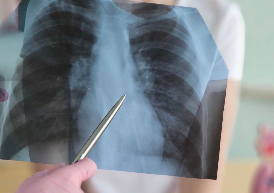
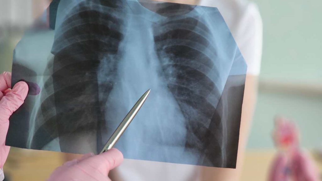
A lung nodule is a small, usually round or oval area seen within the lung on imaging tests. It is often discovered incidentally during a chest X-ray or computed tomography (CT) scan performed for another reason.
Finding a lung nodule does not automatically mean lung cancer. Previous infections, scar tissue, benign tumors, inflammatory conditions, and several other causes may also produce a nodular appearance in the lungs.
However, certain imaging features, growth over time, and individual risk factors may make further evaluation necessary.
The main goal in lung nodule assessment is not to operate on every nodule, but to determine which nodules can be safely monitored and which require additional investigation.
What Is a Lung Nodule?
A lung nodule is a localized area of increased density within the lung tissue. On CT imaging, it appears different from the surrounding normal lung.
Lung nodules may:
- be solitary or multiple,
- be completely solid,
- have a ground-glass appearance,
- contain both solid and ground-glass components,
- remain stable over time,
- or increase in size.
The diameter of the nodule is important, but its structure and changes over time are equally relevant.
What Causes Lung Nodules?
Lung nodules may develop for many different reasons.
Some of the more common causes include:
Previous Infections
Tuberculosis, certain fungal infections, and other previous lung infections may leave small areas of scar tissue after healing.
These areas may appear as nodules on CT scans.
Granulomas
Granulomas are small inflammatory structures that may form as part of the body’s response to certain infections or inflammatory processes.
Many granulomas are benign.
Benign Lung Tumors
Benign tumors such as hamartomas may also appear as lung nodules.
Inflammatory and Rheumatological Conditions
Certain autoimmune, rheumatological, or inflammatory diseases may cause nodular changes in the lungs.
Lung Cancer
Some lung cancers may first appear as a small lung nodule, particularly in the early stages.
For this reason, nodules with suspicious imaging characteristics require careful assessment.
Spread of Cancer from Another Organ
In patients with a previous history of cancer elsewhere in the body, some lung nodules may represent metastatic disease.
Even in this situation, however, imaging alone is not always sufficient to establish a definite diagnosis.
Is a Lung Nodule Dangerous?
Not every lung nodule is dangerous.
A considerable proportion of lung nodules are caused by benign conditions. However, certain features may increase the possibility that a nodule is malignant.
Evaluation usually considers factors such as:
- size of the nodule,
- shape and border characteristics,
- whether it is solid or ground-glass,
- presence or absence of calcification,
- whether it has increased in size,
- location within the lung,
- age of the patient,
- smoking history,
- previous history of cancer,
- personal and family medical history.
Two patients with nodules of the same size may therefore require very different management plans.
Why Is the Size of a Lung Nodule Important?
Nodule size is one of the main factors considered during assessment.
In general, very small nodules tend to have a lower likelihood of malignancy. As a nodule becomes larger or develops certain suspicious imaging features, closer follow-up or further testing may be considered.
However, surgery is never determined by diameter alone.
For example, a small nodule with irregular borders or documented growth may require closer evaluation, while a larger nodule that has remained unchanged for years and has benign imaging characteristics may be managed differently.
Do Lung Nodules Cause Symptoms?
Most lung nodules do not cause symptoms.
They are frequently discovered incidentally during a chest X-ray or CT scan performed for another reason.
In some cases, symptoms may be related to the underlying condition rather than the nodule itself.
These symptoms may include:
- cough,
- shortness of breath,
- chest pain,
- coughing up blood,
- unexplained weight loss.
However, these symptoms are not specific to lung nodules and may occur in many other conditions.
What Happens After a Lung Nodule Is Found?
When a lung nodule is detected, the first step is usually a detailed evaluation of its imaging characteristics and the patient’s clinical background.
Current CT images may also be compared with previous scans.
Determining whether the nodule has remained stable over months or years can provide important information.
Depending on the individual case, evaluation may include the following methods.
Computed Tomography
CT imaging is used to assess the size, shape, density, borders, and location of the nodule in detail.
Comparison with Previous CT Scans
Previous imaging studies can be particularly valuable.
A nodule that has increased in size over time may require a different approach from one that has remained unchanged.
PET-CT
PET-CT may be used in selected cases to evaluate the metabolic activity of a lung nodule.
However, PET-CT cannot by itself confirm cancer.
Inflammatory and infectious conditions may also show increased metabolic activity, while some small or slow-growing cancers may show little or no significant uptake.
Biopsy
Depending on the size and location of the nodule, tissue sampling may be performed through bronchoscopy or image-guided needle biopsy.
Biopsy is not equally suitable for every nodule.
Surgical Biopsy or Resection
In selected cases, surgical removal of the nodule may be considered both for diagnosis and treatment.
When Is a Lung Nodule Monitored Rather Than Treated?
For many low-risk lung nodules, surgery is not the first step.
Instead, the nodule may be monitored with follow-up CT scans at planned intervals.
The timing and duration of follow-up depend on factors such as:
- size of the nodule,
- type and density of the nodule,
- age of the patient,
- smoking history,
- imaging characteristics.
The aim is to avoid unnecessary invasive procedures while still identifying significant changes at an appropriate stage.
What Does Growth of a Lung Nodule Mean?
Growth during follow-up is an important finding.
However, enlargement does not automatically mean cancer.
Some infectious and inflammatory nodules may also change in size.
For this reason, the rate of growth, internal structure, borders, and other imaging characteristics are assessed together.
If the level of concern increases, PET-CT, biopsy, or surgical evaluation may be considered.
When Is a Biopsy Needed for a Lung Nodule?
The decision to perform a biopsy is individualized.
A biopsy may be considered when:
- the nodule has suspicious imaging characteristics,
- growth is detected during follow-up,
- the patient has significant risk factors for lung cancer,
- PET-CT or other imaging findings increase suspicion,
- or a tissue diagnosis is needed to guide management.
Needle biopsy is not always possible or appropriate.
Very small nodules, difficult anatomical locations, or nearby structures may affect both the safety and diagnostic accuracy of the procedure.
When Does a Lung Nodule Require Surgery?
The presence of a lung nodule alone is not an indication for surgery.
Surgical evaluation may become appropriate when:
- the nodule has a high level of suspicion for cancer,
- significant growth is seen during follow-up,
- PET-CT or other imaging findings are concerning,
- biopsy shows cancer or suspicious cells,
- biopsy does not provide a definitive diagnosis but the likelihood of malignancy remains high,
- surgical removal is considered appropriate for both diagnosis and treatment.
Before surgery, the patient’s general health, lung function, and ability to tolerate an operation are also assessed.
Which Surgical Procedures Are Used for Lung Nodules?
The type of surgery depends on the size and location of the nodule, the suspected diagnosis, and the patient’s overall condition.
Wedge Resection
A wedge resection removes a small, wedge-shaped portion of lung tissue containing the nodule.
It may be used for diagnostic purposes or in selected small lesions.
Segmentectomy
A segmentectomy removes one anatomical segment of a lung lobe.
It may be considered in selected early-stage lung cancers or for lesions in suitable locations.
Lobectomy
A lobectomy involves removal of one of the lobes of the lung.
It remains an important surgical option for selected patients with early-stage lung cancer.
The most appropriate operation is determined according to the pathology, location of the lesion, stage of disease, and the patient’s pulmonary reserve.
Can a Lung Nodule Be Removed with Minimally Invasive Surgery?
In suitable patients, lung nodules may be removed using minimally invasive surgical techniques.
Video-assisted thoracoscopic surgery, known as VATS, is performed through small incisions using a camera and specialized surgical instruments rather than a large thoracic incision.
Robotic thoracic surgery may also be used in selected patients.
However, minimally invasive surgery is not suitable for every patient or every type of nodule.
The location and size of the lesion, its relationship with surrounding structures, and the planned extent of resection all influence the choice of surgical approach.
What Happens If a Lung Nodule Is Not Removed?
There is no single answer to this question.
Nodules considered likely to be benign and that remain stable over time may only require follow-up.
On the other hand, delaying the evaluation of a highly suspicious nodule may result in a possible lung cancer being diagnosed at a later stage.
The correct approach is therefore not to operate on every nodule or ignore every nodule, but to choose a management strategy based on the individual level of risk.
Why Is Early Diagnosis Important If a Lung Nodule Is Cancerous?
When lung cancer is detected at an early stage before it has spread beyond the lung, the available treatment options may be significantly different.
Surgery may be an option for selected patients with early-stage lung cancer.
For this reason, appropriate follow-up and timely investigation of suspicious nodules are important.
At the same time, it is equally important not to assume that every lung nodule represents cancer.
Does Everyone with a Lung Nodule Need to See a Thoracic Surgeon?
Evaluation of lung nodules may involve several specialties, including radiology, pulmonology, thoracic surgery, nuclear medicine, and pathology.
Thoracic surgical assessment may become particularly relevant when:
- surgery is being considered,
- the nodule is difficult to biopsy,
- imaging characteristics raise significant concern,
- or removal of the lesion may be required for diagnosis or treatment.
The specialties involved depend on the individual patient’s findings.
Should a Lung Nodule Always Be a Cause for Concern?
No.
Lung nodules may develop for many different reasons, and a substantial number are benign.
However, some nodules can represent an early sign of lung cancer.
The appropriate approach is therefore neither to ignore the finding nor to assume the worst immediately.
A structured assessment based on imaging characteristics, changes over time, and individual risk factors provides the most appropriate basis for further management.
Frequently Asked Questions
1. Does a lung nodule mean I have cancer?
No. Many lung nodules are caused by benign conditions such as previous infections, granulomas, or benign tumors. The likelihood of cancer is assessed according to the nodule’s features and the patient’s individual risk factors.
2. Can a lung nodule disappear on its own?
Some nodules related to infection or inflammation may become smaller or disappear over time. Others may remain unchanged for many years.
3. At what size is a lung nodule considered dangerous?
There is no single size that automatically makes a nodule dangerous. Larger size may increase concern, but shape, density, growth pattern, and individual risk factors are also important.
4. How quickly can a lung nodule grow?
The growth rate depends on the underlying cause. Comparison with previous CT scans is therefore an important part of evaluation.
5. Can a lung nodule cause chest pain?
Small lung nodules usually do not cause pain and are often discovered incidentally.
6. Is PET-CT always necessary for a lung nodule?
No. PET-CT is not required for every nodule. Its use depends on the size, imaging appearance, and estimated level of risk.
7. Does a lung nodule biopsy always provide a definite diagnosis?
Not always. Biopsy can provide important diagnostic information, but obtaining sufficient tissue may be difficult in some nodules depending on their size and location.
8. Can lung nodule surgery be performed with a minimally invasive technique?
Yes, in selected patients. VATS or robotic thoracic surgery may be suitable depending on the location of the nodule and the planned operation.
9. Can non-smokers develop lung nodules?
Yes. Lung nodules are not limited to people who smoke. Infections, inflammation, benign tumors, and other conditions may also cause nodules in non-smokers.
10. How often should a lung nodule be monitored?
There is no universal follow-up interval. The timing depends on the size, type, imaging characteristics, previous changes, and individual risk factors.
Information Note:
This content is intended for general informational purposes only. The evaluation of a lung nodule depends on its size, imaging characteristics, changes on previous scans, and the patient’s medical history. The information provided here does not replace individual medical assessment, diagnosis, or treatment planning. The need for follow-up, biopsy, or surgery should be determined on a case-by-case basis.