What Is COPD? When Is It Considered Advanced Stage?
25/08/2026
What Is COPD Balloon Treatment?
30/08/2026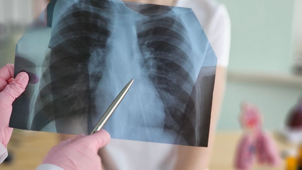
Emphysema is a chronic lung disease that occurs when the air sacs in the lungs, called alveoli, become structurally damaged. Alveoli are small air spaces that allow oxygen to pass into the blood and carbon dioxide to be removed from the body. In emphysema, the walls of these air sacs are damaged, the elastic structure of the lungs decreases, and it becomes harder to move air out of the lungs while breathing out.
Emphysema is usually evaluated together with COPD. COPD stands for chronic obstructive pulmonary disease and refers to a broad disease group characterised by persistent airflow limitation. The main components of COPD may include small airway disease, chronic bronchitis, and emphysema.
Therefore, emphysema and COPD are not exactly the same thing, but they are closely related. Emphysema is one of the structural lung damage patterns that may be seen in people with COPD. In some patients, emphysema is dominant; in others, chronic bronchitis and small airway narrowing are more prominent. In many patients, these findings exist together in different proportions.
When emphysema develops, the lungs may take air in but struggle to let air out. This may lead to air trapping, a feeling of chest expansion, shortness of breath with exertion, reduced exercise capacity, and low oxygen levels in advanced stages.
Emphysema is most commonly associated with cigarette smoke. However, passive smoke exposure, air pollution, occupational dust and chemical exposure, biomass smoke, and genetic causes such as alpha-1 antitrypsin deficiency may also play a role in the development of emphysema.
What Does Emphysema Mean?
Emphysema means permanent damage and enlargement of the alveoli, where gas exchange takes place in the lungs.
Normally, alveoli consist of many small, flexible air sacs. This structure allows oxygen to pass into the blood and carbon dioxide to be removed through breathing.
In emphysema, the alveolar walls are damaged and some small air sacs merge into larger but less functional air spaces.
This may lead to:
-
- Reduced elastic recoil of the lungs
- Difficulty breathing out
- Air trapping in the lungs
- Reduced oxygen transfer into the blood
- Difficulty removing carbon dioxide
- Increased shortness of breath during exertion
- Apparent increase in lung volume
- Greater workload on the respiratory muscles
Emphysema is structural damage with limited reversibility. Treatments do not completely eliminate the disease; they aim to reduce symptoms, slow progression, prevent exacerbations, and improve quality of life.
Why Are Alveoli Important?
Alveoli are the smallest air sacs in the lungs. When we breathe in, air passes through the bronchi into smaller airways and eventually reaches the alveoli.
Very thin blood vessels surround the alveoli. Oxygen passes from the alveoli into the blood. Carbon dioxide in the blood passes into the alveoli and is removed when we breathe out.
Healthy alveoli:
-
- Have thin walls.
- Are flexible.
- Provide a large surface area.
- Make oxygen and carbon dioxide exchange easier.
- Help the lungs empty during exhalation.
When this structure is damaged in emphysema, the problem is not only that the air sacs become enlarged. The gas exchange capacity and elasticity of the lungs are also affected.
For this reason, emphysema is not simply “too much air in the lungs.” The main problem is that air becomes trapped in the wrong place and the lungs cannot work efficiently.
What Happens in the Lungs in Emphysema?
Several main changes develop in the lung tissue in emphysema.
The alveolar walls become damaged. Small air sacs may merge and turn into larger spaces.
The lungs lose elasticity. Normally, after breathing out, the lungs become smaller thanks to their elastic recoil. In emphysema, this elastic recoil decreases.
The small airways collapse more easily during exhalation. This causes air to become trapped inside the lungs.
Lung volume may increase. Because air remains trapped inside, the chest may feel expanded.
Gas exchange may become impaired. Oxygen may pass into the blood less efficiently, and low oxygen levels may develop in advanced stages.
The respiratory muscles work harder. The diaphragm may flatten, and more energy is spent on breathing.
All these changes cause the patient to experience shortness of breath, especially during exertion. In advanced stages, the person may feel breathless even at rest.
Is Emphysema COPD?
Emphysema is closely related to COPD, but it is not the whole of COPD by itself.
COPD is a chronic lung disease characterised by persistent airflow limitation. In COPD, the problem is not only in the alveoli. The small airways, bronchi, and lung tissue may all be affected.
Emphysema is the component of COPD that refers to alveolar damage and destruction of lung tissue.
Simply stated:
-
- COPD is a broad clinical disease definition.
- Emphysema describes structural damage in the lung tissue.
- Chronic bronchitis describes the clinical condition involving cough and sputum.
- Small airway disease refers to narrowing and inflammation in the smallest branches of the bronchi.
A person may have COPD with minimal emphysema. Another person may have marked emphysema with less chronic bronchitis. Some people have both emphysema and chronic bronchitis prominently.
What Is the Relationship Between COPD and Emphysema?
The relationship between COPD and emphysema can be understood through the airway and lung tissue components of the disease.
In COPD, airflow limitation usually develops through a combination of two main mechanisms.
The first is narrowing, inflammation, and mucus increase in the small airways. This makes it harder for air to pass through the bronchi.
The second is lung tissue damage caused by emphysema. When the alveolar walls are destroyed, the lungs lose elasticity and the airways collapse more easily during exhalation.
These two mechanisms work together and make it harder for air to leave the lungs.
The more prominent emphysema is in a patient with COPD:
-
- Air trapping may be greater.
- Shortness of breath may be more noticeable.
- Exercise capacity may be lower.
- Oxygen exchange may be more affected.
- Loss of lung tissue may be seen on CT.
- Volume reduction treatments may be considered in advanced stages.
However, emphysema is not present at the same level in every patient with COPD. Therefore, treatment should be planned according to the patient’s COPD pattern and dominant findings.
What Is the Difference Between Emphysema and Chronic Bronchitis?
Emphysema and chronic bronchitis are two different conditions that are often discussed within COPD.
Emphysema
Emphysema is damage to the alveoli and lung tissue. The main problem is loss of lung elasticity and air trapping.
In emphysema, the following may be more prominent:
-
- Shortness of breath with exertion
- Air trapping
- Hyperinflation of the lungs
- Impaired oxygen exchange
- Feeling of chest expansion
- Weight loss and muscle weakness
Chronic Bronchitis
Chronic bronchitis is characterised by long-term cough and sputum production. There is inflammation and increased mucus production in the inner surface of the bronchi.
In chronic bronchitis, the following may be more prominent:
-
- Morning sputum
- Cough during the day
- Frequent bronchitis episodes
- Chest congestion
- Wheezing
- Increased sputum amount
Can They Occur Together?
Yes. Many patients with COPD have both emphysema and chronic bronchitis features. Therefore, patients are often not divided clearly into “only emphysema” or “only chronic bronchitis.”
The treatment plan is based on the patient’s dominant symptoms, spirometry results, CT findings, exacerbation frequency, and oxygen status.
What Causes Emphysema?
Emphysema may develop when lung tissue is damaged by long-term harmful exposures or genetic susceptibility.
The most important cause is cigarette smoke. Cigarette smoke may cause chronic inflammation, oxidative stress, damage to alveolar walls, and loss of elastic tissue in the lungs.
Causes that may increase the risk of emphysema include:
-
- Smoking
- Passive cigarette smoke
- Pipe, cigar, and other tobacco products
- Air pollution
- Occupational dust exposure
- Chemical fumes
- Occupational environments such as mining, textile work, metal work, and construction
- Biomass smoke
- Heating or cooking with wood, coal, or dung
- Lung development problems during childhood
- Frequent respiratory infections
- Asthma and airway hyperresponsiveness
- Alpha-1 antitrypsin deficiency
- Genetic susceptibility
Not every smoker develops emphysema. However, smoking is one of the strongest and most common risk factors. Likewise, never having smoked does not completely eliminate the possibility of emphysema.
How Does Smoking Cause Emphysema?
Cigarette smoke causes long-term inflammation in the lung tissue.
During this inflammation, the effect of some enzymes that break down lung tissue increases. Normally, the lungs have protective mechanisms against these destructive effects. However, cigarette smoke disrupts this balance.
Over time:
-
- Alveolar walls become thinner and break down.
- Lung elasticity decreases.
- Small airways narrow.
- Mucus production increases.
- Air trapping develops.
- Oxygen exchange becomes impaired.
After smoking cessation, existing emphysema does not completely disappear. However, stopping smoking may slow disease progression, reduce exacerbation risk, and help treatments work more effectively.
Even low levels of smoking may continue to damage lung tissue. For this reason, complete smoking cessation is one of the most important steps in emphysema management.
Can Non-Smokers Develop Emphysema?
Yes. Emphysema may also occur in people who have never smoked.
Non-smoking causes include:
-
- Passive smoke exposure
- Long-term air pollution
- Indoor smoke exposure
- Biomass fuel use
- Occupational dust and chemical exposure
- Alpha-1 antitrypsin deficiency
- Genetic susceptibility
- Previous severe lung infections
- Lung development abnormalities
- Certain connective tissue diseases
Especially in people who develop emphysema at a young age, have no smoking history, or have a family history of similar lung disease, genetic causes such as alpha-1 antitrypsin deficiency should be evaluated.
What Is Alpha-1 Antitrypsin Deficiency?
Alpha-1 antitrypsin is a protein that protects lung tissue from the effects of certain destructive enzymes.
When this protein is deficient or does not function properly, lung tissue may be damaged more easily. As a result, emphysema may develop, especially at a younger age.
Alpha-1 antitrypsin deficiency may be considered when:
-
- Emphysema occurs at a young age
- Emphysema develops in a non-smoker
- There is a family history of early COPD or emphysema
- Emphysema is especially prominent in the lower lung zones
- Liver disease is also present
- COPD cannot be explained by standard risk factors
This condition can be investigated with blood tests and genetic evaluation. If diagnosed, family members may also need assessment.
What Are the Types of Emphysema?
Emphysema can be described in different types according to its distribution in the lung and the structures it affects.
Centriacinar Emphysema
Centriacinar emphysema is most commonly associated with smoking. It may be more prominent in the upper lung regions.
In this type, the areas around the small airways are more affected. It is one of the common emphysema types seen in patients with COPD.
Panacinar Emphysema
In panacinar emphysema, the alveolar structure is affected more diffusely. It may be associated with alpha-1 antitrypsin deficiency.
It may be more prominent in the lower lung regions. This distribution requires particular attention in younger people and in those with little or no smoking history.
Paraseptal Emphysema
Paraseptal emphysema may be seen near the outer edges of the lung. It may sometimes be associated with air spaces called bullae.
In some people, it may be related to pneumothorax, meaning lung collapse.
Bullous Emphysema
In bullous emphysema, large air spaces called bullae form inside the lung. These bullae may compress surrounding healthier lung tissue.
Large bullae may increase shortness of breath in some patients and may require specialised surgical evaluation.
What Are the Symptoms of Emphysema?
Emphysema symptoms may develop gradually. In the early period, the person may notice difficulty only during exertion.
The most common symptoms include:
-
- Shortness of breath with exertion
- Difficulty climbing stairs
- Inability to walk quickly
- Reduced exercise capacity
- Feeling of tightness in the chest
- Wheezing
- Chronic cough
- Sputum
- Need to take deep breaths
- Frequent respiratory infections
- Shortness of breath in cold weather
- Fatigue
In advanced stages, the following symptoms may occur:
-
- Shortness of breath at rest
- Running out of breath while speaking
- Need to stop even after short distances
- Weight loss
- Muscle wasting
- Bluish discolouration
- Low oxygen levels
- Sleep disturbance
- Swelling in the ankles
- Frequent hospital visits
- Slow recovery after exacerbations
Sputum may not always be very prominent in emphysema. It is more common in patients with a dominant chronic bronchitis component.
Why Does Shortness of Breath Occur in Emphysema?
One of the main causes of shortness of breath in emphysema is air trapping.
The patient cannot fully empty the lungs while breathing out. At the next breath, the lungs are still partly full. This leaves less room for new air, and the person feels as though they cannot get enough breath.
In addition:
-
- Lung elasticity decreases.
- The diaphragm flattens.
- Respiratory muscles work harder.
- Oxygen exchange may become impaired.
- Air trapping may increase during exertion.
- Heart and muscle diseases may worsen breathlessness.
For this reason, patients with emphysema often describe the feeling as “I breathe in, but it is not enough.” In many cases, the issue is not only breathing in but also failing to breathe out sufficiently.
Do Cough and Sputum Occur in Emphysema?
They may occur. However, cough and sputum are not present at the same level in every patient with emphysema.
In emphysema-dominant COPD, shortness of breath may be more prominent, while in chronic bronchitis-dominant disease, cough and sputum may be more noticeable.
Cough and sputum may increase when:
-
- Smoking continues
- Chronic bronchitis is present
- There is a respiratory infection
- A COPD exacerbation develops
- Bronchiectasis is present
- Reflux or sinusitis is present
An increase in sputum amount, darkening of sputum colour, clear worsening of shortness of breath, or fever should be evaluated for exacerbation.
How Is Emphysema Diagnosed?
Emphysema is diagnosed by evaluating the patient’s symptoms, risk factors, examination findings, pulmonary function tests, and imaging methods together.
The diagnostic process may include:
-
- Smoking and tobacco use
- Passive smoke exposure
- Occupational dust or chemical history
- Degree of shortness of breath
- Presence of cough and sputum
- Exacerbation history
- Pulmonary function test
- Chest X-ray
- Computed tomography
- Oxygen saturation
- Arterial blood gas
- Diffusing capacity test
- Exercise capacity assessment
- Alpha-1 antitrypsin test
Emphysema is often shown structurally more clearly with computed tomography. However, spirometry is required for a COPD diagnosis because COPD is a clinical and functional disease definition.
Does a Pulmonary Function Test Show Emphysema?
A pulmonary function test helps assess the airflow limitation and lung function loss caused by emphysema.
In spirometry, especially the following are evaluated:
-
- FEV1
- FVC
- FEV1/FVC ratio
For COPD diagnosis, a post-bronchodilator FEV1/FVC ratio below 0.70 is important for persistent airflow limitation.
However, spirometry does not show the distribution of emphysema in the lungs. Computed tomography is more valuable for this.
Some people may have emphysema findings on CT while spirometry does not yet meet COPD diagnostic criteria. In this situation, the patient’s risk factors, symptoms, and follow-up plan should be carefully evaluated.
What Does Computed Tomography Show in Emphysema?
Computed tomography is an important method for assessing the distribution and severity of emphysema in the lungs.
CT may show:
-
- Reduced density in lung tissue
- Areas of alveolar destruction
- Air trapping
- Upper-lobe predominant emphysema
- Lower-lobe predominant emphysema
- Diffuse emphysema
- Bullae
- Bronchial wall thickening
- Bronchiectasis
- Lung nodules or other additional findings
Tomography is especially important in evaluating advanced treatment options. The distribution of emphysema may determine suitability for bronchoscopic valve treatment, lung volume reduction surgery, or bullectomy.
However, tomography is not repeated routinely in every patient. Its need is determined according to the patient’s symptoms and the clinical question.
Can Chest X-Ray Show Emphysema?
Chest X-ray may show some findings of advanced emphysema. However, it is limited in detecting early or mild emphysema.
Chest X-ray may show:
-
- Hyperinflation of the lungs
- Flattening of the diaphragm
- Increased front-to-back chest diameter
- A narrower-looking heart shadow
- Bulla-like areas
- Findings related to other lung diseases
Chest X-ray is not the main test that confirms emphysema. It is mostly used to rule out other diseases or evaluate conditions such as infection, mass, heart enlargement, or lung collapse.
What Is the Diffusing Capacity Test?
The diffusing capacity test helps assess the ability of oxygen to pass from the lungs into the blood.
In emphysema, diffusing capacity may decrease because the alveolar surface area is reduced. This may be especially meaningful in patients whose shortness of breath is more severe than expected from spirometry values.
Low diffusing capacity may:
-
- Support the presence of emphysema
- Show that gas exchange is affected
- Suggest risk of low oxygen during exertion
- Be used in advanced treatment evaluations
- Become important in lung transplantation or volume reduction treatment assessment
However, this test does not diagnose the condition by itself. It should be interpreted together with other tests.
Why Are Oxygen Saturation and Blood Gas Important?
As emphysema progresses, oxygen transfer into the blood may become more difficult. Therefore, oxygen saturation and blood gas evaluation are important.
Oxygen saturation measured from the fingertip may provide a quick idea. However, arterial blood gas may be needed in some situations.
Blood gas can assess:
-
- Oxygen pressure in the blood
- Carbon dioxide level
- Acid-base balance of the blood
- Respiratory failure status
In advanced emphysema, low oxygen levels or high carbon dioxide levels may be seen. These findings affect the treatment plan. Long-term oxygen therapy or non-invasive ventilation may be considered in some patients.
Are There Stages of Emphysema?
There is no simple staging system used universally for emphysema alone. Usually, COPD staging, pulmonary function tests, symptom level, and CT findings are evaluated together.
In COPD, spirometric assessment may be made as GOLD 1, GOLD 2, GOLD 3, and GOLD 4.
For emphysema, the following are important:
-
- Extent of emphysema on CT
- Upper-lobe or lower-lobe distribution
- Homogeneous or heterogeneous pattern
- Presence of bullae
- Degree of air trapping
- FEV1 level
- Diffusing capacity
- Oxygen status
- Exercise capacity
- Exacerbation history
- Daily life impact
Therefore, the answer to “What stage is my emphysema?” should not be based on a single CT sentence. The clinical picture and pulmonary function should be evaluated together.
What Does Mild Emphysema Mean?
Mild emphysema usually means limited lung tissue damage on CT.
In this situation, the person may not have obvious symptoms or may feel breathless only during intense exertion.
However, mild emphysema does not mean unimportant. In a person who continues smoking, the disease may progress.
When mild emphysema is detected:
-
- Smoking should be stopped.
- Passive smoke should be avoided.
- Occupational exposure should be reduced.
- Spirometry should be performed.
- Symptoms should be followed.
- Vaccinations should be considered.
- Exercise habits should be maintained.
- Alpha-1 antitrypsin deficiency should be investigated when necessary.
Correct interventions in the early period may slow progression.
What Does Advanced Emphysema Mean?
Advanced emphysema refers to more widespread or more severe lung tissue damage.
In advanced emphysema:
-
- Shortness of breath may significantly affect daily life.
- Air trapping may increase.
- FEV1 may fall.
- Diffusing capacity may decrease.
- Low oxygen levels may develop.
- Exercise capacity may become limited.
- Weight loss and muscle wasting may occur.
- Exacerbations may be more severe.
- Advanced treatment options may be considered.
Advanced emphysema is not assessed only by the phrase “diffuse emphysema” on CT. Pulmonary function tests, oxygen status, daily functional capacity, and response to treatment should also be considered.
Can Emphysema Progression Be Stopped?
Alveolar damage that has already occurred in emphysema usually cannot be completely reversed. However, the rate of disease progression may be slowed.
The most important steps include:
-
- Complete smoking cessation
- Avoiding passive smoke
- Protection from air pollution
- Reducing occupational dust and chemical exposure
- Correct inhaler treatment
- Vaccinations
- Pulmonary rehabilitation
- Regular exercise
- Early treatment of exacerbations
- Nutritional support
- Special assessment if alpha-1 antitrypsin deficiency is present
- Regular pulmonology follow-up
In emphysema, treatment is not built on erasing existing damage, but on preserving the remaining lung capacity as effectively as possible.
How Is Emphysema Treated?
Emphysema treatment is planned individually. The aim is to reduce symptoms, prevent exacerbations, improve exercise capacity, manage low oxygen levels, and slow disease progression.
Treatment options may include:
-
- Smoking cessation
- Avoiding risk factors
- Inhaler medications
- Pulmonary rehabilitation
- Vaccinations
- Exacerbation treatment
- Long-term oxygen therapy
- Non-invasive ventilation
- Nutritional support
- Exercise programme
- Bronchoscopic volume reduction treatments
- Lung volume reduction surgery
- Bullectomy
- Lung transplantation assessment
The same treatment is not used for every patient with emphysema. Treatment differs between a patient with mild emphysema and a patient with advanced emphysema, low oxygen levels, and daily life limitation.
Why Is Smoking Cessation the Most Important Step in Emphysema?
Smoking cessation is one of the most important steps in emphysema treatment.
When smoking is stopped:
-
- The rate of lung damage progression may decrease.
- Exacerbation risk may decrease.
- Cough and sputum may decrease.
- Exercise capacity may be better preserved.
- Inhaler treatments may work more effectively.
- Lung cancer and heart disease risk may decrease.
- Suitability for surgery or advanced treatments may improve.
Stopping smoking does not completely reverse existing emphysema. However, continuing to smoke may lead to faster loss of remaining lung capacity.
Therefore, the thought “the damage is already done” is misleading. Damage may already exist, but preserving the remaining lung tissue is still essential.
Do Inhaler Medications Cure Emphysema?
Inhaler medications do not eliminate the alveolar damage caused by emphysema. However, they may reduce shortness of breath and make daily life easier by widening the airways.
Inhaler medications that may be used include:
-
- Short-acting bronchodilators
- Long-acting bronchodilators
- Dual bronchodilator combinations
- Combinations containing inhaled corticosteroids depending on exacerbation risk
Treatment selection depends on the patient’s symptoms, FEV1 value, exacerbation history, blood eosinophil level, accompanying asthma features, and previous treatment response.
Correct inhaler technique is very important. If the inhaler is used incorrectly, the medication may not reach the lungs.
Does Pulmonary Rehabilitation Help in Emphysema?
Yes. Pulmonary rehabilitation is one of the most important parts of emphysema and COPD treatment.
Pulmonary rehabilitation programmes may include:
-
- Exercise training
- Breathing techniques
- Energy conservation methods
- Disease education
- Inhaler technique training
- Nutritional support
- Psychological support
- Exacerbation awareness
Patients with emphysema may avoid movement because of shortness of breath. Inactivity increases muscle loss. As muscles weaken, the person becomes tired more quickly and shortness of breath becomes more noticeable.
Pulmonary rehabilitation may help break this cycle. Even if it does not completely eliminate breathlessness, it may help the patient move more safely, maintain muscle strength, and remain more independent in daily life.
When Is Oxygen Therapy Needed in Emphysema?
Oxygen therapy is not given to every patient with emphysema. Shortness of breath alone is not enough to require oxygen use.
Long-term oxygen therapy is considered in selected patients with persistent low oxygen levels at rest.
Oxygen therapy may be considered when:
-
- Oxygen level is low at rest
- Oxygen pressure in blood gas is below a defined threshold
- Low oxygen has increased strain on the heart
- Pulmonary hypertension has developed
- Signs of right heart failure are present
- Significant oxygen drop occurs during exertion or sleep
Oxygen therapy should be planned by a physician. Oxygen equipment should not be used randomly, because oxygen is a medical treatment, not an ordinary household device.
Why Are Vaccinations Important in Emphysema?
Respiratory infections may trigger exacerbations in patients with emphysema and COPD. Exacerbations may worsen lung function, increase the risk of hospitalisation, and make recovery more difficult.
For this reason, suitable patients may be evaluated for:
-
- Influenza vaccine
- Pneumococcal vaccine
- COVID-19 vaccine
- RSV vaccine
- Other vaccines appropriate for age and risk status
Vaccinations do not treat emphysema. However, they may help reduce infection-related worsening.
What Is an Exacerbation in Emphysema?
An exacerbation is a clear worsening of the patient’s respiratory symptoms beyond normal daily variation.
During an exacerbation:
-
- Shortness of breath may increase.
- Cough may increase.
- Sputum amount may increase.
- Sputum colour may darken.
- Wheezing may increase.
- Fever and weakness may occur.
- Oxygen may drop.
- Emergency care may be needed.
- Hospitalisation may be needed.
Exacerbations are important in the course of emphysema and COPD. Treatment should be reassessed in patients who have frequent exacerbations.
Recognising exacerbation symptoms early is important for starting treatment in time and preventing unnecessary worsening.
What Is Bronchoscopic Valve Treatment in Emphysema?
Bronchoscopic valve treatment is a volume reduction method that may be considered in selected patients with advanced emphysema.
In this method, one-way valves are placed into the airways using bronchoscopy. The aim is to reduce the air volume of an overinflated and poorly functioning lung region and help healthier lung areas work more effectively.
However, this treatment is not suitable for every patient with emphysema.
Suitability assessment considers:
-
- Distribution of emphysema
- Whether it is heterogeneous or homogeneous
- Fissure integrity
- Presence of collateral ventilation
- FEV1
- Lung volumes
- Diffusing capacity
- Exercise capacity
- Exacerbation history
- Oxygen status
- General health status
Valve treatment is evaluated in advanced centres and selected patients. It is not a simple process of “there is emphysema, so a valve should be placed.”
What Is Lung Volume Reduction Surgery?
Lung volume reduction surgery is the surgical removal or reduction of the most damaged and overinflated lung regions in advanced emphysema.
The aim is to create space for healthier or better-functioning lung regions, help the diaphragm work more effectively, and reduce shortness of breath.
This method may be considered especially in selected patients with advanced emphysema and certain distribution patterns.
Assessment includes:
-
- Distribution of emphysema
- Pulmonary function tests
- Exercise capacity
- Oxygen and carbon dioxide status
- Heart health
- General surgical risk
- Pulmonary rehabilitation status
- Smoking cessation status
This surgery is not performed in every patient with advanced emphysema. Incorrect patient selection may bring more risk than benefit.
What Is Bullectomy?
In bullous emphysema, large air spaces may form inside the lungs. These spaces may compress surrounding healthier lung tissue.
Bullectomy aims to remove large bullae that cause compression.
This procedure may be considered especially when:
-
- A large bulla compresses surrounding lung tissue
- It increases shortness of breath
- There is a risk of recurrent pneumothorax
- Infection or bleeding develops
- There is enough functional lung tissue around it
Not every bulla requires surgery. Small bullae that do not cause symptoms may be followed.
Is Lung Transplantation Considered in Emphysema?
In very advanced emphysema or advanced COPD, lung transplantation may be considered in selected patients.
However, an emphysema diagnosis alone is not enough for lung transplantation.
Assessment includes:
-
- Age
- General health status
- Heart function
- Oxygen need
- High carbon dioxide levels
- Exacerbation frequency
- Exercise capacity
- Weight and muscle status
- Smoking cessation status
- Treatment adherence
- Infection risk
- Cancer history
- Psychosocial support
Lung transplantation is not suitable for every patient with advanced emphysema. Suitable patients are evaluated in detail by transplant centres.
Does Emphysema Increase Lung Cancer Risk?
Emphysema requires attention in terms of lung cancer risk, especially when combined with a history of smoking. The presence of emphysema on computed tomography may be an important finding in lung cancer risk assessment.
In people with emphysema, the following should be evaluated together:
-
- Smoking history
- Age
- Pack-year calculation
- Family history
- Presence of lung nodules
- Previous CT findings
- Occupational exposure
In suitable risk groups, lung cancer screening with low-dose computed tomography may be considered. However, the decision for screening should be based on individual risk assessment.
Does Emphysema on CT Always Mean COPD?
No. Seeing emphysema findings on CT does not always mean that a spirometric COPD diagnosis has been made.
Some people may have areas of emphysema on CT, while their pulmonary function test does not yet show an FEV1/FVC ratio below the diagnostic threshold for COPD.
In this situation, the person should not be considered completely normal, because there is structural lung change. However, COPD diagnosis requires clinical assessment and spirometry.
In such a case:
-
- Smoking should be stopped.
- Risk factors should be reduced.
- Spirometry should be performed.
- Symptoms should be followed.
- Pulmonology assessment should be performed when necessary.
- Lung cancer screening suitability should be evaluated according to risk.
A single CT phrase should not be treated as destiny, but it should also not be ignored. A balanced approach is needed.
Can Emphysema Be Confused with Asthma?
Yes. Emphysema, COPD, and asthma may overlap in terms of symptoms.
All three conditions may cause:
-
- Shortness of breath
- Wheezing
- Cough
- Chest tightness
- Difficulty with exertion
Asthma usually has a more variable course. Symptoms may occur in attacks, may be related to allergy, and may show more obvious improvement after bronchodilator use in pulmonary function testing.
COPD and emphysema are more associated with persistent airflow limitation. A history of smoking or harmful gas-dust exposure is more common.
Some patients may have features of both asthma and COPD. Therefore, treatment planning should be based on detailed assessment rather than a single symptom.
Are Emphysema and Pulmonary Fibrosis the Same Thing?
No. Emphysema and pulmonary fibrosis are different diseases.
In emphysema, alveolar walls are destroyed and air spaces increase in the lung tissue. The lungs may become overinflated and lose elasticity.
In pulmonary fibrosis, hardening and scar tissue develop in the lung tissue. The lungs have difficulty expanding, and a restrictive type of pulmonary function disorder may occur.
In some patients, emphysema and fibrosis may exist together. This situation requires more complex assessment.
Computed tomography and pulmonary function tests are important in making this distinction.
Can Emphysema and Bronchiectasis Occur Together?
Yes. Emphysema and bronchiectasis may coexist in some patients.
Bronchiectasis is permanent widening and structural damage of the bronchi. It may present with frequent infections, sputum, cough, and exacerbations.
If bronchiectasis is present together with emphysema:
-
- Sputum may be greater.
- Infection risk may increase.
- Exacerbations may be more frequent.
- Treatment planning may change.
- Sputum culture may be needed.
- Airway clearance methods may become important.
Therefore, on CT, not only emphysema but also accompanying bronchiectasis, nodules, fibrosis, or other findings should be assessed carefully.
What Happens When Emphysema Is Considered Advanced?
When emphysema is considered advanced, the disease needs closer follow-up and wider evaluation of treatment options.
In advanced disease, the following may be assessed:
-
- Suitability of inhaler treatment
- Inhaler technique
- Exacerbation prevention
- Pulmonary rehabilitation
- Oxygen need
- Blood gas evaluation
- Exercise capacity
- Nutritional status
- Muscle strength
- Heart and pulmonary vascular pressure
- Suitability for bronchoscopic valves
- Suitability for lung volume reduction surgery
- Lung transplantation assessment
- Palliative care and symptom control
Advanced stage does not mean “there is nothing left to do.” That would be both medically incomplete and unnecessarily discouraging for the patient.
How Does Emphysema Affect Daily Life?
Emphysema especially affects exercise capacity.
Over time, the patient may:
-
- Need to stop while climbing stairs.
- Be unable to walk quickly.
- Struggle when walking uphill.
- Become breathless while carrying shopping.
- Need breaks during housework.
- Become tired while bathing or dressing.
- Reduce social activities.
- Avoid going outside.
- Develop sleep problems.
- Experience anxiety or panic.
As shortness of breath increases, the person avoids movement. As movement decreases, muscles weaken. As muscles weaken, shortness of breath becomes more noticeable. Pulmonary rehabilitation and regular safe movement are important to break this cycle.
What Should Patients with Emphysema Pay Attention To?
Daily life adjustments are an important part of treatment in people diagnosed with emphysema.
Important points include:
-
- Smoking should be stopped completely.
- Passive smoke should be avoided.
- Dust, smoke, and strong odours should be avoided.
- Inhaler medications should be used with correct technique.
- Follow-up appointments should not be missed.
- Vaccinations should be assessed.
- Regular physical activity should be maintained.
- Pulmonary rehabilitation should be followed if recommended.
- Nutritional support should be obtained if there is malnutrition.
- Exacerbation symptoms should be recognised early.
- Oxygen therapy, if recommended, should be used correctly.
- Indoor air quality should be improved.
- Care should be taken in cold weather and during periods of heavy air pollution.
Emphysema follow-up is not just about receiving medication. Treatment, lifestyle, and monitoring should be carried out together.
When Is Emergency Care Needed in Emphysema?
Some symptoms in patients with emphysema or COPD may require urgent assessment.
Emergency care is needed when there is:
-
- Sudden increase in shortness of breath
- Breathlessness at rest
- Bluish lips or fingers
- Confusion
- Excessive drowsiness
- Inability to speak because of breathlessness
- Chest pain
- Blood in sputum
- High fever
- Clear change in sputum amount and colour
- Significant drop in oxygen saturation
- Worsening despite home medications
- Sudden sharp chest pain with shortness of breath
- Feeling faint
Especially in people with bullous emphysema, sudden chest pain and shortness of breath should also be evaluated for pneumothorax.
Which Doctor Should Be Consulted for Emphysema?
The main specialty for emphysema diagnosis and follow-up is pulmonology.
Initial assessment may be performed by a family physician or internal medicine specialist. However, pulmonology assessment is important for pulmonary function testing, CT evaluation, inhaler treatment, oxygen need, pulmonary rehabilitation, and advanced treatment options.
In advanced treatments, the following specialties may also be involved:
-
- Thoracic surgery
- Interventional pulmonology
- Cardiology
- Physical medicine and rehabilitation
- Nutrition and dietetics
- Intensive care
- Lung transplant centre
- Palliative care
The patient’s treatment plan should be made according to the whole clinical picture, not a single test result.
Conclusion
Emphysema is a chronic lung disease that develops when the alveoli in the lungs are damaged and the lungs lose elasticity. Because of this damage, it becomes harder to breathe air out of the lungs, air trapping develops, and shortness of breath may become more noticeable over time.
Emphysema and COPD are not the same thing. COPD is a broad disease definition characterised by persistent airflow limitation. Emphysema is the lung tissue damage component that may be seen within COPD. Chronic bronchitis, small airway disease, and emphysema may coexist in different proportions.
The most common cause of emphysema is cigarette smoke. However, emphysema may also develop in non-smokers due to passive smoke, air pollution, biomass smoke, occupational exposures, and alpha-1 antitrypsin deficiency.
Diagnosis involves pulmonary function testing, computed tomography, oxygen measurement, blood gas analysis, diffusing capacity, and assessment of risk factors. Spirometry is important for COPD diagnosis, while computed tomography provides more detailed information about the structural distribution of emphysema.
Treatment may include smoking cessation, inhaler medications, pulmonary rehabilitation, vaccinations, prevention of exacerbations, oxygen therapy, and, in advanced patients, bronchoscopic or surgical volume reduction procedures. In very advanced and carefully selected patients, lung transplantation may be evaluated.
Emphysema is not a fully reversible disease. However, with correct follow-up and treatment, progression can be slowed, shortness of breath can be reduced, exacerbations can be prevented, and quality of life can be preserved.
Frequently Asked Questions About Emphysema
1. What Is Emphysema?
Emphysema is a chronic lung disease in which the air sacs called alveoli are damaged and lung elasticity decreases. It becomes harder to move air out while breathing out.
2. Is Emphysema COPD?
Emphysema is closely related to COPD, but it is not the whole of COPD. COPD is a broad disease definition; emphysema is the lung tissue damage component that may be seen within COPD.
3. What Is the Difference Between Emphysema and Chronic Bronchitis?
In emphysema, alveolar damage and air trapping are prominent. In chronic bronchitis, long-term cough and sputum production are more noticeable. Many patients with COPD may have both.
4. What Are the Symptoms of Emphysema?
The most common symptoms are shortness of breath with exertion, easy fatigue, wheezing, chest tightness, cough, and reduced exercise capacity. In advanced stages, shortness of breath at rest, low oxygen, and weight loss may occur.
5. How Is Emphysema Diagnosed?
Diagnosis includes pulmonary function testing, computed tomography, oxygen saturation, blood gas analysis, diffusing capacity, and assessment of risk factors. Spirometry is the main test for COPD diagnosis.
6. Does Emphysema on CT Mean COPD?
Not always. Emphysema may be seen on CT, but spirometry may not yet meet COPD diagnostic criteria. In this situation, pulmonology assessment and follow-up are needed.
7. Can Non-Smokers Develop Emphysema?
Yes. Passive smoke, air pollution, occupational dust and chemical exposure, biomass smoke, and alpha-1 antitrypsin deficiency may cause emphysema in people who have never smoked.
8. Can Emphysema Be Completely Cured?
Existing alveolar damage usually does not fully reverse. However, smoking cessation, correct treatment, pulmonary rehabilitation, and prevention of exacerbations may slow disease progression.
9. Are Valves or Surgery Used in Emphysema?
Bronchoscopic valve treatment, lung volume reduction surgery, or bullectomy may be considered in selected patients with advanced emphysema. These treatments are not suitable for every patient and require detailed assessment.
10. Which Doctor Should Be Consulted for Emphysema?
A pulmonologist should be consulted for emphysema diagnosis and follow-up. For advanced treatment options, thoracic surgery, interventional pulmonology, rehabilitation, cardiology, or transplant centre evaluation may be needed.